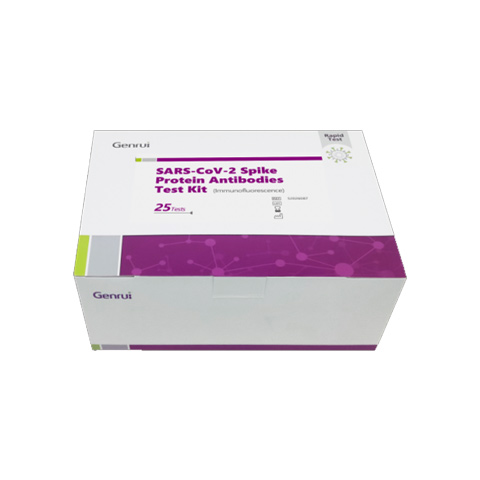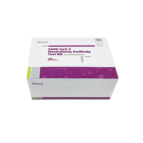
Immunofluorescence antibody test is the immunoassay technique that uses a detector antibody or an antigen labeled with florescent dyes.
Immunofluorescence (IF) is a common morphological approach used to determine the distribution of subcellular components. Immunofluorescence antibodies that conjugated with fluorescent dyes are required in IF assay. The antibody specifically recognizes the antigen by binding to the epitope of target, and the fluorophore will be detected under a fluorescent microscope. Hence, subcellular components can be visualized in a dark background. IF can also be used as an alternative semiquantitative analysis method to monitoring the expression of the interest.

Clear positioning. This assay can give research on the clear subcellular localization of molecules. The expression of molecules can be observed directly.
High specificity. The preparation of the sample can effectively protect the natural structure of the antigen
High sensitivity. The target with low expression can be detected with immunofluorescence.
Easy to operate. The test result can get quickly. This assay can be used for in-vivo experiments.
Multiple staining. The multiple targets can be detected in the same sample.
Immunofluorescence is an assay which is used primarily on biological samples and is classically defined as a procedure to detect antigens in cellular contexts using antibodies. The specificity of antibodies to their antigen is the base for immunofluorescence.
Fluorescent dyes (fluorochrome) illuminated by UV lights are used to show the specific combination of an antigen with its antibody. The antigen-antibody complexes are seen fluorescing against a dark background.
Direct FAT is used to detect and identify an unknown antigen in specimens, for example, Viral, bacterial, and parasitic antigens.
It is called direct test because the fluorescent dye is attached, or labeled, directly to the antibody.
The fluorochrome used is usually fluorescein isothiocyanate (FITC), which gives a yellow-green fluorescence.
A fluorescent substance is one that, when absorbing light of one wavelength, emits light of another (longer) wavelength.
In the indirect FAT, the unlabelled antibody combines with antigen and the antigen-antibody complex is detected by attaching a fluorescent-labeled anti species globulin to the antibody.
The antibody, therefore, is labeled indirectly. Fluorescent-labeled antihuman globulin is used if the antibody is of human origin.
 10
10Feb.10, 2025
Live From Dubai: Genrui at Medlab Middle East 2025 15
15Nov.15, 2024
Live From Düsseldorf: Genrui at Medica 2024 02
02Aug.02, 2024
Live From Chicago: Genrui at ADLM 2024

